Compact Bone Diagram Microscope - Mammal Compact Bone, c.s. 7 m H&E Microscope Slide: Microscope Sample Slides: Amazon.com .... Hematoxylin & eosin decalcified bone. Compact bone specimens (2x2x36 mm) were harvested from the right metatarsal. A diagram of the anatomy of a bone, showing the compact bone. Microscopic examination of compact bone, however, reveals that it is riddled with passageways 4 2. Compact bone consists of closely packed osteons or haversian systems.
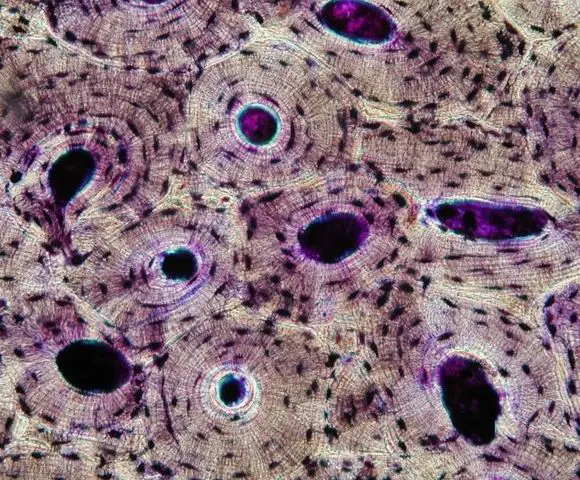
Decalcified compact bone at 60x magnification. As seen in the image compact bone is formed from a number of osteons, which are circular units of bone material and blood vessels. The osteon consists of a central canal called the osteonic (haversian) canal, which is surrounded by concentric rings (lamellae) of matrix. These units allow compact bone to. They consist of two outer layers of compact bone and an inner layer.
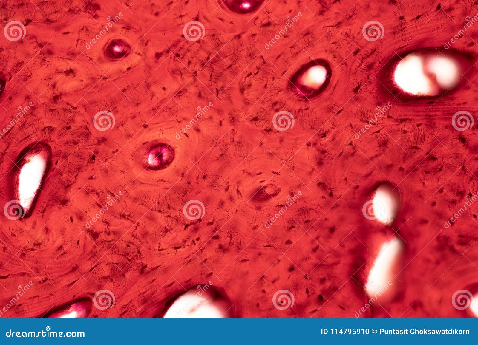
Supporting Connective Tissue | Human Anatomy and Physiology Lab (BSB 141) from s3-us-west-2.amazonaws.com In this type of bone, the lamellae are organised into concentric circles, which surround a vertical haversian canal (which transmits small neurovascular and lymphatic vessels). Decalcified compact bone at 60x magnification. They consist of two outer layers of compact bone and an inner layer. It's easy to look at these and think of bones as dry, dead sticks in your body, but this couldn't be further from the truth. The series of diagrams below represent the microscopic structure of compact bone tissue. The ground substance of bone is arranged in concentrated layers (lamellae) round the small canals which run parallel to the long axis (shaft) of the bone. There are pores and spaces even in compact bone. Compact bone specimens (2x2x36 mm) were harvested from the right metatarsal.
Study flashcards on bones at cram.com.
Because of the short depth of field, students should have no problem focusing on. Learn vocabulary, terms, and more with flashcards, games, and other study tools. Electron microscope image of trabecular bone (x100 magnification). Samples were cyclically loaded to failure and then histological analyses on each hysteretic diagram, all cycles after the initial monotonic cycle appear pinched and share two points. Most think that bone is a dead tissue, but this is not the case. Compact bone labeled microscopic structure of compact bone labeled compact bone diagram compact bone diagram labeled anatomy human. Except at its edge, the osseous tissue of compact bone is arranged. Compact bone, also known as cortical bone, is a denser material used to create much of the hard structure of the skeleton. A diagram of the anatomy of a bone, showing the compact bone. Zeiss microscopy is a leader in microscopy for biomedical and materials research this week, we introduced zeiss primostar 3. In this type of bone, the lamellae are organised into concentric circles, which surround a vertical haversian canal (which transmits small neurovascular and lymphatic vessels). If you look at compact bone under the microscope, you will observe a highly organized arrangement of concentric circles that look like tree trunks. Migliaia di nuove immagini di alta qualità aggiunte ogni giorno.
Zeiss microscopy is a leader in microscopy for biomedical and materials research this week, we introduced zeiss primostar 3. Most think that bone is a dead tissue, but this is not the case. 2 compact bone we know that compact bone is very dense it is also very complex when viewed under a microscope. Quickly memorize the terms, phrases and much more. Electron microscope image of trabecular bone (x100 magnification).
Bone Tissue and Cells Under The Microscope from www.microscopemaster.com Two structural arrangements of bone tissue are seen: Start studying microscope structure of compact bone. Between the rings of matrix, the bone cells (osteocytes) are located in spaces called lacunae. Learn vocabulary, terms and more with flashcards, games and other study tools. 3 mature bone cells, osteocytes, are found in 7 bone formation, growth and remodeling the skeleton is formed from two of the strongest and most supportive tissues in the body. Samples were cyclically loaded to failure and then histological analyses on each hysteretic diagram, all cycles after the initial monotonic cycle appear pinched and share two points. A cross section of decalcified compact bone is examined under brightfield illumination with the intel qx3 microscope. These units allow compact bone to.
Samples were cyclically loaded to failure and then histological analyses on each hysteretic diagram, all cycles after the initial monotonic cycle appear pinched and share two points.
Under the microscope dense, compact bone shows a definite and a characteristic pattern of arrangement. Compact bone diagram bone cross section diagram file624 diagram of compact bone new. Each group of concentric circles (each tree) makes up the microscopic structural unit of compact bone called an osteon (this is also called a haversian. Nov diagram for.net is a fully managed, extensible and powerful diagramming framework, which can help you create feature rich diagramming solutions in winforms, wpf, silverlight, xamarin.mac, monomac and asp. The series of diagrams below represent the microscopic structure of compact bone tissue. Usually bones that are thin and curved. What are the 2 main types of bone? Most think that bone is a dead tissue, but this is not the case. 2 compact bone we know that compact bone is very dense it is also very complex when viewed under a microscope. Start studying compact bone under microscope. Trova immagini stock hd a tema compact bone structure under microscope e milioni di altre foto, illustrazioni e contenuti vettoriali stock royalty free nella vasta raccolta di shutterstock. It's easy to look at these and think of bones as dry, dead sticks in your body, but this couldn't be further from the truth. Because of the short depth of field, students should have no problem focusing on.
Two structural arrangements of bone tissue are seen: 7 m h&e microscope slide. Compact bone diagram osteon compact bone ap pinterest anatomy human anatomy and. Compact bone diagram bone cross section diagram file624 diagram of compact bone new. Histology of human tissue, show skin as seen under the microscope.
Histology Of Human Compact Bone Tissue Under Microscope View For Stock Photo - Image of organ ... from thumbs.dreamstime.com As seen in the image compact bone is formed from a number of osteons, which are circular units of bone material and blood vessels. Study flashcards on bones at cram.com. Between the rings of matrix, the bone cells (osteocytes) are located in spaces called lacunae. Histology of human tissue, show skin as seen under the microscope. Migliaia di nuove immagini di alta qualità aggiunte ogni giorno. A diagram of the anatomy of a bone, showing the compact bone. Learn vocabulary, terms and more with flashcards, games and other study tools. Begin by identifying the concentric rings of lamellar bone that surround a haversian canal.
In this type of bone, the lamellae are organised into concentric circles, which surround a vertical haversian canal (which transmits small neurovascular and lymphatic vessels).
Compact bone, also known as cortical bone, is a denser material used to create much of the hard structure of the skeleton. Learn vocabulary, terms and more with flashcards, games and other study tools. There are pores and spaces even in compact bone. Study flashcards on bones at cram.com. This item mammal compact bone, c.s. Electron microscope image of trabecular bone (x100 magnification). Most think that bone is a dead tissue, but this is not the case. 7 m h&e microscope slide. Each group of concentric circles (each tree) makes up the microscopic structural unit of compact bone called an osteon (this is also called a haversian. What are the 2 main types of bone? Compact bone labeled microscopic structure of compact bone labeled compact bone diagram compact bone diagram labeled anatomy human. The pictures on this card were take through a microscope under low power (100x) and under high power. Histology of human tissue, show skin as seen under the microscope.
The transmitted brightfield digital images above were recorded using a qx3 microscope that was modified for auxiliary illumination compact bone diagram. Under the microscope, bone can be divided into two types compact bone forms the outer 'shell' of bone.
Share :
Post a Comment
for "Compact Bone Diagram Microscope - Mammal Compact Bone, c.s. 7 m H&E Microscope Slide: Microscope Sample Slides: Amazon.com ..."


Post a Comment for "Compact Bone Diagram Microscope - Mammal Compact Bone, c.s. 7 m H&E Microscope Slide: Microscope Sample Slides: Amazon.com ..."